


So we can probably agree that it is not host specific, or at least a braodleaf parasite with minimal damage to the host
Beiträge von Steve_mt
-
-
Here I found this species (unless it was a different but closely related one) on Eucalyptus!!. On oaks too and apparently in Malta prefers carob trees.
-
Hello again - PCR and sequencing were successful on persisting a bit more with Pablo (he is really a good guy) and I have a name for this collection. I don't know if it is actually it or a very close relative (new species) but the answer is Clavaria salentina (a recent description from Sicily). It looks alike and a bit different too.
Clavaria salentina - A.M.B. PESAROGenere Clavaria Specie salentina Agnello & Baglivo, 2010 Posizione sistematica: Ordine Agaricales, Famiglia Clavariaceae. Exsic. n. 201 Erbario Maletti.…www.ambpesaro.itQuel manto arancione presso una masseria identificata come nuova specie di fungoMESAGNE – La sua caratteristica colorazione arancione, contribuisce a fare del variopinto mondo delle biodiversità, comprese nella macchia mediterranea, un…www.brindisireport.itI look forward to contribute more to this lovely community
-
I also think it is an infection from a parasitic fungus, not 'own' mycelium.
 Alles anzeigen
Hello Timm,
is that really mycelium?
Reminds me of a yoke mushroom:
Syzygites megalocarpus,
it affects other fungi too.
At least you can compare that.
VG
Thomas
-
I see
 so probably the mould has caused the spores to have that sunken shape ... though to be fair, I have not seen (at least evidently) alien hypha when viewing the sporocarps under the microscope. Maybe I get new growth because I have saved the plant-cutting (Centaurea ragusina) in a jar of water, re moisted the old leaves and let it to stand/dry maybe some spores will germinate and I have fresh specimens. Many Thanks Ulla
 -
https://www.discoverlife.org/mp/20q?search=Perichaena+depressa
Oh my - yes I think you are correct. It is a beautiful myxo, hope I have fresh regrowth to photograph fresh specimens, maybe they grow a bit larger too.
Thank you!
-
Hope you can confirm with me that this Arcyria cinerea. Cysts on stipe were observed.
-
Hello,
Here is another Physarum on decaying plants which I am trying to identify. I only have about 15 fruiting bodies (maybe more will grow) and they are old I think. The myxocarps are very rich in lime almost fluffy, the spores are like collapsed but few were examinable and are round to slightly oval, not ornamented and unless it is my imagination, i am seeing a faint line across some. Those which are starting to collapse or shrivel show this line stronger (line of weakness ?). The lime nodules are chunky and quite rounded at edges. Spores 11-12 um (so it fits too). I am sorry that I do not have better material!
-
Hello, I found some myxomycetes on a plant with dead leaves immersed in water, and this tiny species a discoid myxocarp with orange spores mass is one of them. The fruiting body is minute barely visible and I have very little material to examine. The spores seems be ornamented with very small spines (punctate) and the capillitium is very thin and wavy. Some spores have an equatorial marking (maybe an artefect).
pilzforum.eu/attachment/426960/
-
Good afternoon and good Friday.
Today I spent some time checking some moulds that I have grown from random sources, including contaminants in previous isolations, air-borne spores, etc. More or less to self-educate. The one I am posting about should correspond to a Botrytis sp. Colonies are pale greyish with a hint of olive-brown hue or greenish in LED light. AT first, I thought I was examining B. cinerea, the common botrytis, but on consulting some books, the species is likely to be B. aclada or B. allii (the paper below recognizes both species as valid) and both are distributed worldwide and closely related to each other. The spore size may tell them apart.
Conidiospores were measured and have the following results:
(6.5) 7.2 - 9.5 (9.8) × (3.1) 4 - 4.9 (5.4) µm
Q = (1.5) 1.7 - 2.1 (2.5) ; N=39
V = (39) 64 - 110 (153) µm3
Me = 8.5 × 4.4 µm ; Qe = 1.9 ; Ve = 89 µm3
According to the article below the small conidiospores correspond to B. aclada!
Chilvers, MI, and du Toit, LJ 2006. Detection and identification of Botrytis species associated with neck rot, scape blight, and umbel blight of onion. On-line. Plant Health Progress doi:10.1094/PHP-2006-1127-01-DG.
Download paper (Chilvers, & du Toit, 2006)
I wonder if my analysis is correct and this species should correspond to B. alcada.
My thanks
-
Hi, I am also studying some Cosmospors/Stylonectria which were growing on pyrenomycetous fungi on the bark of various trees. Seems it is not an easy road, as first, we have to isolate the fungus on media, study the anamorph and then we can narrow down to species. Your pics are fantastic btw!
-
Thanks bjorne - I had my browser set to - automatic translate to english and the chapters of the book were showing in English on Springer website. Pity the book is in German. Wish it will get translated soon or later as it seems to be a great reference and unique book
(maybe if I buy the pdf where I can copy text and get the text block translated in Google translate.... but not sure if the pdf let u to copy text to be pasted (sometimes they don't allow it for copyright)thanks
-
Hello Bjorn! Thanks for the detailed reply and it was very helpful.
Starting from the last, the book should be this one:
Pflanzenparasitische Kleinpilze | SpringerLinkMaybe I buy the e-book at half the price.
Regards our rust, I scraped immediately my idea of P. striiformis and it is most likely to be Uromyces lineolatus. From the aspect of host ecology (life cycle) there are several Apiaceae in the surroundings (Daucus, Ferula and Phoeniculum) while there are no Nymphoides recorded in Malta (and cultivated chances has to be discarded as the locality is away from urban). I can mount some spores in glycerol solution and see if they germinate to have the 100% mark.
The 1959 book is here (german text
)THANKS
-
Uromyces lineolatus | (Obligat) Phytoparasitische Kleinpilze
Uromyces lineolatus ?
"this rust fungus performs a host change. While the spermogonia and aecia are formed on umbelliferae (Apiaceae), the fungus switches to sea rush ( Bolboschoenus ) in summer for the formation of uredia and telia ."
-
This is a Pucciniaforming pockets elongated from where the spores emerge and released. I think Identification of Puccinia is somewhat host-related. I don't know if there is a website/database where one can input the host and get a set of possible plant pathogens.
Spores
(23.6) 24.7 - 32.1 (32.5) × (16.4) 17.2 - 19.8 (20.1) µm
Q = (1.2) 1.25 - 1.8 (1.9) ; N=9
V = (4018) 4518 - 5293 (5427) µm3
Me = 27.1 × 18.6 µm ; Qe = 1.5 ; Ve = 4892 µm3
and spore wall is almost 2 um thick!
Well if you have some information to share with me about this species I will be glad.
pilzforum.eu/attachment/426295/pilzforum.eu/attachment/426296/
-
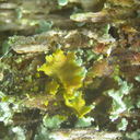
Hello guys... I am really busy concluding my thesis (on macromycetes) of which deadline is in a few weeks now. I think many remember this post and its mysterious orange coral-like fungus, where I sent samples for DNA analysis. Few days ago I received the DNA ITS1 results and disappointingly Dr Alvarado told me it was noisy or contaminated and suggested to repeat. I have the fasta file (I can share with whome is interested to compare) but maybe its wise to wait the repeat. I am quite sure I did not contaminate the specimens.
-
-
Thanks for replying Thorben. Ok we exclude Trichoderma. When I am not busy I try to match it graphically or try to key it out. I posted it because it is quite beautiful.
-
I wonder if this is another Trichoderma sp., Different from the one I recently posted.
This is also a contaminant while trying to isolate Cosmospora from organic substrate on culture media
-
Tirchoderma is a good option here or there are other genera please ? The perpendicular profuse branching and fluffy-green colonies do match.
-
Thanks, I selected this mold for sequencing - I keep u posted - hope it works out.
1. Dried agar at 35-39 C
2. Cut agar into small pieces (quarters)
3. Placed in an aluminum pocket
4. Then in a paper pouch
5. Placed paper pouched in a bag with silica gel
P..s. in the agar I added antibiotics to prevent bacterial growths (it helps a lot) hope this would not interfere with the sequencing!
-
Thanks, yes. Not very human-friendly pet!
Thanks
-
Some more data
Colonies had a strangeoff-putting rubbery scent (I think I should not have smelled it!)
On applying KOH the spores turns dull brown
The conidiophore os smooth, thin
The heads are two-series, the metulae elongated trapezoid, the phialides short
Metulae 20-28 um long
Phialides 7-8 um long
Spores 3-4 um across, smooth
-
According to my simple book (but I love its keys) this would be A. flavus (easy peasy as that ? I don't think so !) but I am seeing colonies of A. parasiticus offer a closer match!
-
Here are some microscopical data.
The heads are in my opinion une-seriate, but better confirm from the images. The spores are spinulose, 5-6 um, they do not change colour in KOH. The conidiophores are thick and widen close to the head and there is some 'wrinkling ridges' just below the head (characteristic ???) and covered with dense small warts. Phialides are about 12-15 um long with tapering tips. Conidiospores are produced in long chains and are difficult to separate from the heads. The heads are quite strong, I could not break one to get loose the phialides!