


I do that tomorrow
Beiträge von Steve_mt
-
-
I got that idea because I can see light (= hollow / depressed centre). Also the large-sized pileus (15 cm across) triggered that species, yet I just suggested without any solid determination.
-
At first, I thought this is some small Gomphidius but it lacks a ring (ring scar), pileus not moist or slimy and the stipe is a bit different too. I defaulted to Clitocybe (nebularis?) but I am not saying that is the genus, just logic thoughts. The two specimens were ca. 1.5 cm across and short.
-
Are they really decurrent or raised at an angle due to the orientation of the pileus? ( kinda a bit funnel-shaped) ?
-
Is it Coriolopsis gallica please? I have a sample at home. The hispid upper surface, habitat and colours of the Hymenium suggest this species.
-
Hello Germany. I hope you can help me with this exciting finding growing on wet soil covered with moss.
I have collected a sample (including soil) and stored it in the fridge so as I want to have advice from you on how to proceed to determine the genus (and species!).
I noticed a pruinosity on the surface, when slightly touched, the outer surface easily breaks in releasing its sap and hence the pruinosity islost leaving a darker color (hope I explained myself well here). There seams to be a stipe that is yellowish. Verry irregular fruiting bodies. It is not in my opinion: Leotia, Auricularia or Dacrymyces (but D. palmata looks a bit similar, but this is a wood saprotroph).
If these photos can't tell the species I am interested to learn how to carry on. I have three (four) specimens.
-
I has similarities with Megacollybia platyphylla -
 Alles anzeigen
 Alles anzeigen
Eckelig, EVERYONE
Oh, the green Knolli still looks delicious == Gnolm7
And if the Steinis weren't bad, they'd probably be good too.
And with the stinker, the tastiest thing is already gone.
A suitable method of preparation would only have to be invented for the "snails".
Unfortunately nothing pissed off from me and Irisle ...Greetings from Ali
Wutzi has already got a message from me
As rotten as it is, I will never touch it!
Yeah... I don't like to eat left overs too 😅
-
We don't have those Russulas 😭😭😭 Can I apply to some chemical or compound instead? Or fabric or ironmongery material.
-
I have acquired an old bottle with Guaiac reagent which was evaporated in its reagent bottle (10-15 mls). I added alcohol and the reagent dissolved forming a brown solution without precipitate. I don't know if this solution is working (reacting) well or if it has 'expired', hence how can I test it at home pls? Like I am asking how can I test for Lugol's iodine and the reply would be try it on starch or paper and if it turns blackish-blue it is working fine. Thanks
-
I have concluded upon M. olivaceomarginata based on the text by Aronsen fabolous website:
One should also notice a possible confusion [of M. capilleripes] with Mycena olivaceomarginata (Massee) Massee, which sometimes has a greyish brown pileus with a reddish brown center as well as a stipe with a dingy whitish apex and greyish brown colors below, and a nitrous smell. The two species can be told apart on account of the cheilocystidia. In M. capillaripes they are smooth while they are more varied in M. olivaceomarginata , often with two or three necks or with several coarse excrescences. The Latter therefore lacks pleurocystidia.
Moreover, Mycena olivaceomarginata has been reported and associated with Cistus matorral (specifically C. monspeliensis) while M. capilleripes is associated with pine forests. Loizides 2016, 2021
-
Hi, are here some mycenologists? I have found two interesting Mycena species (I am working / documenting one of them here) which was found under Cistus monspeliensis. The species has striking lilac then rosy-pink gill edges hence a species from the section Rubromarginatae.
I can't decide between
Mycena capillaripes and M. olivaceomarginata
I read somewhere that olivaceomarginata was recorded under cistus monspeliensis too (Lozoides)
-
They should be more or less soft when young and crumbly when decaying.
Lg; pablo.
Yes, that's what the case and also dark reddish-brown when overmature and decaying out. I think I found a similar specimen on Celtis australis again above grown and on the bark of a living tree on mainland Malta. Crumbling brown debris was at the foot of the trunk and fall down on the ground
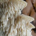
ZitatThey are really ennoying under the microscope! Hard, dense - difficult to prepare and difficult to interprete.: gkopfkratz:
Yes, I haven't grasped very well the concept of monomitic / dimitic / trimitic, generative hyphae, skeletal hypha, etc when examining under the microscope. They just look like a brush of unspecialised mycelia entangled into each other. I had a look again this morning and I an interesting parasite!
-
Wow! ...and I was sure it was a Panaeolus sp. for its marbled face and white edge of the gills. So are there more genera which are Panaeolus-like and have these characters ??? Fungi are very confusing!
Axel and Karl - I think you are bull's eye regards your kind suggested identity. The pic is identical! Tonight or tmrw I compare and show you the spores. Their hexagon-like thick outline seems quite unique. THANK YOU again.
Deconica coprophila ( syn. Psilocybe coprophila ) aka the dung-loving psilocybe (lol!)
-
I am linking to a post o the main forum topic in case some dung-specialists only check this corner.
Follow the link below.
I was surprised that when I search for "fimicola", there were no results. Is it not reported from Germany / central Europe?
Steve
-
Very interesting and tiny mushroom. I can't help just wanted to say impressive. Hope you get it identified and I think it is rare finding. good job
-
I forgot to include a few more photos of when it is hygrophanous dry. I guess i need to offer the microscopy for this one... but let's see if I get some feedback based on the macro-morphology
-
Thank you so much xxx
Take care everyone, love this group!
Steve
-
A Coprinoid fungus on horse dung, highly hygrophanous, semi-spherical pileus, caramel brown with a beige rim when wet, then whiter and zonated when dey. I thought of Panaeolus fimicola. Thank in advance for your kind comments on this.A sample is in the fruidges
Cheers
-
Nice fungus and widespread population on horse dung sitting amongst a grassy lawn. I have identified it from the macro aspect as Panaelous papilionaceus s.l. (the length of the spores will tell the var). Hope it is OK.!!!
L..G.
Stephen / Gozo (Malta)
-
In the first photo on the left I think that is a decaying example which gave me the guidance of being an annual polypore.
I can monitor regularly (this is a park near to work) and see what happens when they mature. If they decompose and die (=annual) it might be I. dryophila. They were attached to bark and even cortex of living holm oak trees
I try to provide images of the context. So if I see septation that's a big clue ?
-
OK, I trust you guys. I try and slice the other one although its older. I show you the spores too tomorrow or Tuesday
THANKS!
-
It is matching quite will with Parasola leiocephala = P. lactea .
12. Basidiospores narrowly ovoid to ellipsoidal, sometimes
ovoid, Qavg = 1.15–1.60 ................................ 13
12′. Basidiospores ovoid, rounded triangular, or subglobose
...................................................................... 14
13. Basidiospores mostly ellipsoidal to hexagonal, Qavg
= 1.15–1.5, pleurocystidia mostly
utriform ....................................................... P. plicatilis
13′. Basidiospores broadly ellipsoidal, often ovoid to
broadly hexagonal, Qavg = 1.25–1.6, pleurocystidia
often lageniform .......................... P. plicatilis-similis
14. Average length of basidiospores under 10.5–11 μm,
basidiospores often remain immature
.................................................. P. lactea (= P. leiocephala
14′. Average length of basidiospores >11.5 μm, as usual
fully mature, dark blackish brown, pileipellis and
basidia filled with strongly refringent, yellowish
granules ................................................P. lilatincta s.l.
-
Thank you Tuppie... You are right about the colour, but I cant find a better option for my very very limited knowledge on boletes. We see what Beorn opines

Happy Sunday to all
Stephen
-
And yet another Xerocomus under Quercus ilex in a park. No signs of reddening in the context of the entire stipe and pileus. No blueining on touching the pores or cut the basidiocarp. There is a scent of something but I cant tell, like aromatic. Basidiocarps quite large 9 cm the largest one. Is X. subtomentosus a good option here. I have half the specimen in the desiccator and half for the spore print / fresh. Habitat match nicely - oaks in parks!