Hello, everyone, and I hope you had a good start for 2025.
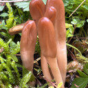
I encountered a Tricholoma sp. with a dark reddish-brown cap , white gills which turn cream or pale brown on aging (due to spore cover) , and a white stipe which turns light beige with time. Spore print chocolate brown. Several specimens were under Pine or Cypress trees in a calcareous ground. I made the mistake of not smelling it!
The spores are quite large with a distinct apiculum, elliptical-phaseoliform, brown.
(10.9) 11.6 - 13 (14.5) × (5.6) 5.9 - 7 (7.6) µm
Q = (1.7) 1.8 - 2 (2.2) ; N = 39
V = (190) 221 - 334 (402) µm3
Me = 12.3 × 6.5 µm; Qe = 1.9 ; Ve = 274 µm3
The cheilocystidia are numerous, narrowly utriform (capitate?) with many horn-shaped crystals at the top.
Pileipellis with dark pigments on the walls. I saw clamp junctions too.
Basidia broad, rounded apex, (2)-4 sporous.
Do I have enough information to give me a clue about the identity?