


Here is another unidentified small pale mushroom with contrasting scales on the pileus and dark punctations on the stipe. I think there was/is a ring but got damaged or eroded away. Pileipellis a trichoderm. Clamp junctions were observed. In a phrygana associated with Cistus monspeliensi (or less likely Thymus capitatus). I was guessing about Lepiota, but which species I don't know. In Crete, Lozoides and allies records a number of fungi associated with Cistus, amongst which there is Lepiota locquinii, L. farinolens and L. sublaevigata.
Beiträge von Steve_mt
-
-
Just a question - Are all Asterophora species parasitic ?
-
Happy birthday Pablo and thanks for the previous helping posts to my ID of fungi.
Hope you had a good day
LG
Steve

-
: D
 love the answer!
 -
Yes, in an upset mood !
-
No.1 I opt for Agaricus section minores for the small size, no yellowing, simple ring.
Then you need microscopical examination, scent and spore sizes...
(PDF) New data about Agaricus (Section Minores, Agaricaceae) in Bulgaria
-
Leucoagaricus maybe? I presume the gills did not darken.
I. familiar with L. leucothites , compare with it, as it looks a bit alike and it is a first-fruiting species in the year. But from photos only, other species can be considered (and some white mushrooms are toxic!)
-
Same thought of LG Ulla (Hygrocybe 100%) though I would say conica s.l. as there is a small group of closely related species (e.g. H. singeri). The vividly coloured Hygrocybe will darken like soot when old.
-
If u can handle Spanish:
Externer Inhalt www.youtube.comInhalte von externen Seiten werden ohne deine Zustimmung nicht automatisch geladen und angezeigt.Durch die Aktivierung der externen Inhalte erklärst du dich damit einverstanden, dass personenbezogene Daten an Drittplattformen übermittelt werden. Mehr Informationen dazu haben wir in unserer Datenschutzerklärung zur Verfügung gestellt.The one in the video is sordida (smaller, thinner, less robust, little/weak violet hues)
B.t look also here
-
Lovely photos gosh! Tubaria 100% and I also opt for T. conspersa. T. confragosa is darker and I would exclude safely
-
Hi, I am no expert, but have you considered/compared with Peziza vesiculosa?
The blisters, colour, size, and irregular shape fits
-
I don't know, but is there a bit of similarity with Asterophora?!?!
Asterophora parasitica / Astérophore parasite | Récolté par … | Flickr
Image - Asterophora parasitica (Silky Piggyback) | BioLib.cz
Parasitic Asterophora - Asterophora parasitica | Björn S... | Flickr
-
Hi, I have checked again the lamellae using a slightly different mounting technique. Lots of details has been preserved, and I can surely see basidia, and spores that are immensely variable in size and abundant. There are also some hyphae that are possibly cheilo/pleuro cystidia. I post everything here!
-
-
Uwe and others who are interested in this fungus, I had time to carry out the examination of the pileipellis. Many parts did not preserve well, but I could see a trichoderm of wavy or almost straight cylindrical hyphae, hyaline, with terminal cells having a very obtuse or truncate ending, and I could not see any clamp junctions.
thats strange, your sure you measured the spores with 400x and not with 1000x ???
I have no idea which white fungus has such large spores.
BR
UweYes, I was also puzzled. I had in mind this strange thought of some sort of Asterophora and we are seeing Chlamydospores ?!?! However this is a parasitic genus right?. I can redo the spores but I am absolutely sure I was under x400. Spores from a contaminant fungus during dehydration is also possible, but again... low possibility.
-
-
Here I am back after finding the ex-siccatum and examined again the spores more carefully under Melzer and Baral Iodine (a stronger version of Lugol's)
1. I think they are inamyloid

2. The spore size is larger than in the first examination, and they average 15.5 x 7.5um.
This large size of the spore might be helpful. Are we still in Clitocybe with these large spores? What about Lyophyllum (Ossicaulis or other genera?) Which like to form fused stipes!
An example of Lyophyllum with chalky pileus
-
Hi, I did not on that occasion, but I have one from another foray from the same place:
-
Marasmius corbariensis, decaying leaves of olive tree in a damp area, 8/10/2021, Malta (Gozo)
My first encounter of a fungus for season 2021-2022 after 5 months of very little rain. The season begins
-
Thank you UWE. Agreed - I was reading about the species and its spores has amyloid ornamentation. == Mushroom25
-
Thank you,
I also have an update (to be confirmed) that I marked on my XL that I saved a ex-siccatum specimen. Maybe it is useless if they collapse but worth a try. I check your post
-
I include this example with 5 spore measurements only so that I keep things simple.
As you see the smallest spore is 6.9 and the largest is 8.2 yet Piximetre ranges the spore sizes 6.2-8.7 and I think the extra 1um (0.5um per end) (c. 18%) is a bit too much erroneous. Should it be something like 6.9 [7.1; 7.7] 8.2 ?!?! I never thought about but when I saw this I was wondering....
6.93 4.87 7.08 5.15 7.89 5.37 7.12 5.58 8.18 5.26 6.2 [6.9; 8] 8.7 × 4.7 [5; 5.5] 5.8 err Q = 1.2 [1.3; 1.5] 1.6; N = 5; C = 95% Me = 7.4 × 5.2 err; Qe = 1.4 -
Thanks Wolfgang. I remember it did not sporulate and when I carried out the pileipillis test I was careful to extract the outermost layer. My fault that I did not carried out a good job on the micro (Images crap!)
It us a very strange species and this year I go and search it again (know the place to go) and follow the tape-method which I like and usually I use it on Penicillium spp. and moulds! Would a diluted drop of Phloxin or Congo red help ?
-
Second opinion is Hygrophorus pseudodiscoideus var. Cistophilus Bon & G. Riousset 1988
But not matching very well in my opinion
-
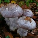
If there are experts who study leucopaxillus / Clitocybe I have found this interesting species Under Cistus monspeliensis, here in Malta. I have tentatively identified it as Leucopaxillus paradoxus for its gross physical characters (robust habit, browning areas in pileus, thick stem, dry chamois-like texture, taste above-average bitter, lamella cream-caramel) and somewhat the micro too: Cheilocystidia not observed, spores large about
7-8 µm15.5 x 7.5 um. Unfortunately, I had not much time to work on it as I collected other fungi which I gave more priority. I read that this species was recorded under pine and oak ... so maybe it is host-variable. The appearance is very matching.