


It is a petiole (stalk) of a leaf of Rubus ulmifolius, but I have also collected the species from legume of decaying carobs
regards Steve!
It is a petiole (stalk) of a leaf of Rubus ulmifolius, but I have also collected the species from legume of decaying carobs
regards Steve!


The Axioscope 20 should be infinite, is it?
Yes, it is!
Sorry folks - My microcope is AxioLab RE not Axioscope 20
The Axioscope 20 should be infinite, is it?
Yes, it is!
Sorry folks - My microcope is AxioLab RE not Axioscope 20
The other old x100 oil eyepiece behaves the same. In oil mount 100um gives 85 reticule units and was shown in the photo.
This is a Zeiss NeoFluar 100\ 1.30 Oel 160\- code 48600960
Please not that my Microscope Is AxioLAB RE
This is the one I am testing right now:
ZEISS Plan-NeoFluar
100x / 1,30 Oil
1018-595 (1066-987)
on the other side
ZEISS Plan-NeoFluar
100x / 1,30 Oil
∞ 0,17
I took photos by digital camera from the eyepiece.
Daturamyces ![]()
What is the best site to order/preview the book please. Sounds appealing to me.
Would be nice if someone can test the same procedure and calibrate the measurements with oil and without oil for the x100 objective and see if there are same results. Because if one measures spores under oil immersion and applies a 1:1 scale, there might be some 15% error in the measurements!!!! --- or is it just me here with this anomaly ?!?!
this morning tried the calibration without oil and I got 96-97 units for 100um (everything same setup but no oil on stage). The focusing and sharpness was quite good actually! That makes 100/97 = 1:1.04 which is a good match.
Replies:
I second Peters opinion that the problem is the measuring scale in the eye piece. >> Same eyepiece reticule for all objectives (which gives expected values)
You should have the same unusual multiplication factors also in the other magnifications, means 1:4,25 with the 40x objective (instead of 1:2,5) and 1:17 with the 10x objective (instead of 1:10) - correct? >>> For the other 'air' objectivesI get a close value to the expected magnification.
Your eyepieces are 10x?
Yes E-Pl 10
Or you can buy a "real microscope" from Olympus right away.![]()
You need real money too!
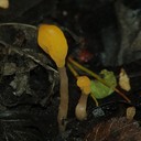
hilmgridd , dont worry about the English, what's important is that we communicate .... (although I couldn't get the joke about the egg lol!). I attach two more photos of this Diderma species from a previous collection which was more typical (well-developed stipe) for you to admire. Yes it is a lovely slime mold looking like tiny satellite radar discs.
Agreed eith LG Ulla, the species is not specific to Juglans (I found it earlier on carob leaves and Rubus ulmifolius decaying stalks) but I think it requires a lot of humidity and likely a thick layer of leaf detritus.
 Alles anzeigen
Dear Steve,
my knowledge about this optical problems is limited. But I will transfer this problem to the microscopy forum, which usually provides very good help. It usually is in German language, but I will send you the results.
all the best,
Andreas
Thanks - but have I posted in the microscopy section already? I start thinking what happens without oil immersion ...and also if users here calibrate the magnification (for measuring) compensated for oil immersion! ! !
Hi there,
I don't really believe that the lens delivers such a clear change in the image scale.
- Was it really readable 1: 1 before?
No, always 1:1.17 (oil mount)
- Is the measuring eyepiece the same as before?
Eyepiece original of microscope (Zeiss Axioscop 20) but reticule different.
- Is the measuring scale in the eyepiece the same? (There are so many different scales)
yes
- Has the graticule slipped in the measuring eyepiece?
No
- Is the microscope micrometer inserted the right way round?
\yes, if not the numbers of the micrometer will be inverted and I would know
The easiest way is to screw in the two 100 lenses side by side and compare them directly.
Peter, maybe you are misinformed, but the lens always gave this 1:1.17, that is, it was not like it was giving 1:1 and now all of a sudden 1.1.17. If I remember the previous x100 objective did the same.
regards
Peter
I wonder if you guys have your x100 objective giving 1:1 or 1:>1? when using oil (that is you put a drop of oil on stage micrometer)
Alles anzeigenHello Steve,
that is the objective from FluorescensMicroscops.com, isn't it?
You have an infinity objective, and unlike the 160 tube length objectives the infinity ones often do only fit in the very microscope version it was made for. So you can normally not use an infinity objective from Zeiss in a microscope from Olympus, just as an example. What microscope do you have and which tube length does it have? I bought a Zeiss 80x infinity objectiv for dry use and tried it on my Olympus BX40 - no chance, doesn't work, bad luck ......
Another idea is that this is probably a objective for fluorescens microscopy and may be it works only properly with a fluorescens equipement?
Oil with wrong refraction index will result in unsharp view, but doen't affect the viewed magnification (at least not that much), and neither does a movement of the object.
all the best,
Andreas
Hi Andreas, I bought it from ebay and there is one like it here:
Zeiss Plan-NEOFLUAR 100x/1.30 Oil Objective
I was told infinity objectives made by Zeiss should work on Zeiss microscopes so I had a budget and this objective looked really good. However I am a bit disappointed with the resolution, and often the lens has to touch the cover slip to have focus. I don't know if, If it does work well for transmitted light application.
Maybe u r right re used ONLY for fluorescent microscopes, but websites selling the exact obj are saying that it could be used both for light and florescent applications ! As far as I know, such a specific lens should be colour coded . where black it means for transmitted light
Zeiss Plan-NEOFLUAR 100x/1.30 Oil Objective
I also have this objective : Zeiss Plan Neofluar 100x/1.30 Oil Pol Microscope Objective Lens 440483 for sale online | eBay
Alles anzeigenHello Steve,
that is the objective from FluorescensMicroscops.com, isn't it?
You have an infinity objective, and unlike the 160 tube length objectives the infinity ones often do only fit in the very microscope version it was made for. So you can normally not use an infinity objective from Zeiss in a microscope from Olympus, just as an example. What microscope do you have and which tube length does it have? I bought a Zeiss 80x infinity objectiv for dry use and tried it on my Olympus BX40 - no chance, doesn't work, bad luck ......
Another idea is that this is probably a objective for fluorescens microscopy and may be it works only properly with a fluorescens equipement?
Oil with wrong refraction index will result in unsharp view, but doen't affect the viewed magnification (at least not that much), and neither does a movement of the object.
all the best,
Andreas
P.s. I am using a Zeiss Axioskop 20 similar to this
 Alles anzeigen
Hi there,
just do a bit physics. The nominal magnification is valid for an exactly positioned object and environment. Whenever you move the object here and there, or adjust the ocular to your eyes - the magnification is also slightly changed. This is unavoidable. And yes, the different refraction index of oil in comparison to air contributes as well.
Much appreciated you checked this!
Best regards,
Bernd
But then the 630x objective at same conditions (except not oil) does not suffer from any discrepancy.
Alles anzeigenHello fungi,
I am always thrilled when a newbie starts this forum with a request for LBMs, ie the "Little Brown Mushrooms", affectionately also known as the "Little Browns"! Seasoned mycologists often need years before they dare to approach these difficult to identify fungi!
Hats off! == Pilz24
Pablo has already said everything about the possible species.
LG, Nobi
nobi_†: So true, but then a genus name is already very good for newbies .
@ Fungi : Agree with the above, there are quite some Psathyrellaceae in the bunch. For better expectations, ideally you post species pre post and take several pics from different angles including scent. In several other fora or social media you want be helped from such a photo.
Thanks! On examining carefully, some had short stipes. Maybe the sudden climate change (warmth in car) and then home resulted in a slightly abnormal formation. Many thanks!
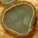
I found an interesting myxomycete, rather abundant on fallen leaves of Juglans regia and few other exotic trees. Initially, they looked like small flour sacks or pouches, quite large (2mm perhaps), and quite cute. On further development, there were some slimy droplets oozing (maybe damaged during transport) and finally, they solidified into irregular flattened disks and dehisced in a particular way. Must be a Diderma sp. and maybe globosum or more likely D. hemisphaericum. However, the latter should be distinctly stalked! On careful examination, the myxocarps were subsessile (neither distinctly stipitate nor sessile). For me, it looks more like hemisphaericum unless there are other species that I have not considered. I am aware micro is needed but for this I might be lucky to decrease the workload!
I was about to write that 3g/100mL = 3% is too concentrated (usually literature suggests 1%). I opt for 0.8% in my preparation and see if it works well.
And just for fun, I found a thesis on colours of Congo Red written in 1927 by Robert S. Radcliffe.
RICE1273.pdf?sequence=1&isAllowed=y
Thanks Andreas and Andreas
Thank you Andreas, what % is the Ammonia, around 10% maybe?
I am preparing new solution from the powder now, that Congo Red is strange.
I take the long cut, I dissect and put the section in congo red. Leave for a minute or so, then I absorb the extra stain with a cotton bud or kitchen roll strips and add plenty of 3%KOH and wash the dissection in it (sometimes leave it to stand for another minute if the tissue is robust/hard). Then I pick the washed section and place it in a drop of KOH+Glycerol mixture, sometimes a dissect further in smaller pieces and space them out. I place the cover slip over, press with a rubber over tissue paper and ready to go. The glycerol mixture prevents the slide from drying.
Cheers ![]()
The one on the right is Congo Red Powder C.I. 22120 dissolved in water and filtered (I asked my friend who gave it to me) and remains red in KOH. Actually, when searching for my stuff, I just realised that I have bought a bottle and forgot about it - so at least this post was very beneficial !!! The one on the left was bought a few months ago from a commercial Lab Supplies and they provided it as a solution (1%). Well, I can make my own Congo Red solution from 22120... I guess 1% is a good concentration or a bit less is better? Since my Lab is home-based I prefer to avoid Ammonia.
P.s. Does it really expires 🤷♀️😁 ?
Hi guys, tomorrow I show you a video of what's happening. Thank you so much for your replies. I am a bit more relaxed with regards toxicity. The other (old) congo red do form some minute crystals and I know the notorious feeling under the microscope. Sometimes it goes away by heating it in water bath at 70C until water cools down gradually
I have had a stock of Congo Red donated by a friend and recently I bought a new stock from a local Lab supplies shop. The second one is a different kind!
In alkaline solution, it becomes purple and somewhat leaches out stain when a stained section is placed in clean KOH.
Are there different types of Congo Red because I may buy again this time from abroad.
Another point, it is really toxic and carcinogenic? Sometimes my fingers are stained and then I nibble my nails! : blow: