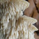
I am just working on some Mycena / Hemimycena specimens!!! That's great share. Thanks!!!
Beiträge von Steve_mt
-
-
hi Steve,
Have you checked amyloidity?
regards Pit
No unfortunately, but my heart tells me it is a member of the Phloeomana group (they are all inamyloid if I remmeber well). Still, I am not convinced it is P. hiemalis (best match) or alba, because there are some considerable mismatches. I discarded the option M. polyadelpha too. Those inflated hyphae are very peculiar!
-
rare occurrence of caulocystidia
-
Some time ago I have found a cluster of small Mycena growing on decaying bramble sticks (Rubus ulmifolius) having a purely white and slightly transparent colour (esp. the stipe), fading a bit into beige when old. Cap 7-12 mm wide with non-numerous, spaced (not crowded) lamellae, horizontally-arched not particularly decurrent. Basidia 4-sporous (sometimes 2). There were swollen hyphae from sub-globose to broadly cylindrical, sometimes with a short foot (sphaeropedunculate) which I think were cheilo or/and pleuro cystidia (40-90 x 25-50 um). Spores 8-10 x 4.5-6 um (Q 1.5-1.9) ovate-elliptical to moderately broadly ovate, very rare to find in slides. I didn't check well the pileipellis. The stipe was more or less glabrous but I think I got a slide showing some caulocystidia.
Do you have any suggestions?
I shortlisted to Hemimycena gracilis, Mycena alba, Mycena pseudolactea, Mycene spiraea
-
Hello Steve,
A. vervacti seems to be impossible. Spores should be 7-9(-10) x 4.5-6 μm
Another species with large spores is A. ochracea with pleurozystidia.
LG KarlThank you Karl. I am more confident about A. pedaides which is frequent in the Mediterranean region.
LG
Steve (Malta)
-
I have an old collection which I initially determined as Agrocybe pediades but now I have a doubt on A. vervacti. It seems both share the same habitat and are not very different macro-morphologically. I am seeking advice from some expert here, maybe there are ways to differentiate the two species. I did not write notes about the smell, unfortunately, but the spores are large and should fit A. pediades better.
I have other microscopical images and measurements.
Spores :
spore Spore length: range 11 - 14.8 µm Spore length: mean 13.2 µm Spore width: range 7.1 - 9.5 µm Spore width: mean 8.5 µm Spore Q factor: range 1.4 - 1.8 Spore Q factor: mean 1.6 Spore shape Elliptical, often symmetrical in both axis (fusiform) Amyloid reaction Inamyloid Spore surface Smooth A little bee Present but inconspicuous Oil bodies Huge and conspicuous singular oil body. Remarks Not congophilous and weak cyanophilous. Germination pore evident. -
For those who are following this thread, after considering everything, I am labelling this collection as C. truncicola
The woody habitat, smaller sporocarp, no conspicuous rhizoids at the base of the stem, and somewhat broader spores (amongst other things) contributed to this decission. C. rivulosa is a grass-loving species which has rhizoids at the stipe, more densely pruinose and overall whitish and tends to be medium-sized (my specimen ca. 2 cm across, 3.5cm tall).
The congophilous reaction on the spores mentioned by Flora Agaricina Need.was interesting and I wonder if it really useful for determining clitocybe sp. (in this case this sp was not congophilous and hence do not stain much with Congo Red)
-
I bumped into this species: Clitocybe truncicola, let's see how much it matches...
Clitocybe truncicola - A.M.B. PESAROGenere Clitocybe Specie truncicola (Peck) Sacc. Posizione sistematica: Ordine Agaricales, Famiglia Tricholomataceae. Sottogenere Disciformis, Sezione…www.ambpesaro.itTrunk funnel (Clitocybe truncicola) - mushrooms of Eastern Texas
-
Yes I know, so I am doing my best to document what I am finding with my limited resources. I often send samples for ITS sequencing but I have to limit the samples as much as possible because I pay everything myself. For Clitocybe, I am using Flora Agaricina Neerlandica Vol3 - I dont' know if there are better / updated sources on the genus.
For this one in particular:
1. Hygrophanous: YES
2. Has pruinosity (aeroferous covering sensu Flora Agaricina Need.) yes but limited at the margin ? border line yes/no
3. Spore print white / Lamella White
4. Spores 4-5.5 um, broadly ovoid
5. Smell indistinct (taste bitter)
5. Lamella very crowded ? I don't know!
6. Stipe short? Yes
Following the Key,
if we consider the specimen pruinose it would key best as C. rivulosa
if we consider the specimen pruinosity limited to the margin, it would key best as C. diatreta (although this is said to have a cream spore print).
Reading further FAN3 , it says that C. diatreta has spores that are congophilous whereas C. rivulosa are not congophilous (as were in this specimen). Spore size matching slightly better rivulosa too. The habitat of rivulosa is grassy but sometimes on wood, that of dealbata is more woody in mossy or grassy ground, so the habitat goes a bit in favour to diatreta.... but overall within the species list and descriptions of the FAN3 key, I see it more rivulosa. If there are other species to consider, I don't know!
-
Found attached to decaying twigs of a carob tree in a shaded and humid environment. Spore print white. No particular odour, taste bitter, unpleasant.
I was thinking on Clitocybe rivulosa but this grows on grass.
I haven't see cheilocystidia (if present rare and not detected)
Spore Details:
Spore length: range 3.9 - 5.7 µm Spore length: mean 4.6 µm Spore width: range 2.9 - 4.2 µm Spore width: mean 3.4 µm Spore Q factor: range 1.2 - 1.8 Spore Q factor: mean 1.4 Spore shape Elliptical with a small apiculus Amyloid reaction Inamyloid, (and doubtfully mildly dextrinose) Spore surface Smooth of finely rough texture (few wide, plug-shaped and wart-like projections observed but are considered as artifacts) Apiculum Present and conspicuous Oil bodies Oil bodies not conspicuous Remarks Weak stain in congo red, hence spores not congophilous -
I want to discuss something I noticed today while cleaning my microscope desk and lenses.
I have two ocular micrometres of different tube lengths. So I, while checking and experimenting on the stereomicroscope, I discovered something which made me wonder and worry.
Using the same subject (letter R of a receipt), I put the adjustable eyepiece setting of the stereomicroscope all way up and then all way down and realised that the length of the 'R' varied from 16 to 20 divisions (c. 25% difference). I repeated this with the other micrometer. R measured differently between 19 to 23 divisions - four different readings for the same subject!!!. I was not aware that this would have any effect (never been told during courses on microscopy). It may be a potential source of error when measuring spores in the light microscope if the microscope eyepiece tube goes up and down (not the lens itself)
So this makes calibration a bit more awkward and the following steps should be adopted:
1. Put the micrometer always on the same eypiece and set it fixed in middle way setting (o).
2. Focus compensation between eyepieces should hence be done on the other eyepiece only
3. Using this fixed setting, perform callibration for each micrometer
I thought this is good to share and discuss!
-
How much would you sell the x63 lens (i do not require the trinocular) Also what brand and characteristics does the lens bear?
-
Hi Ingo.
Thanks for sharing your experience. Would freezing samples for a day kill the eggs/larvae and leave the fungal tissue good for microscopy and molecular analysis later? Can this be adapted to to some larvae of maggots in soft mushrooms ? ( who feast out when drying specimens)!
BTW I couldn't go on the link provided - would love to have a look.
In this case the best thing to do is ask and send samples to an expert, if he is interested to follow up in the material. I do have some bugs in 70% ethanol and of course a stereo microscope.
Cheers
-
This is a bit off topic but I think it is both related and educative.
I was cleaning my mycology desk and preparing for a fresh start for the next season, and there were some old specimens of bracket fungi (Inonotus and Ganoderma, etc ). The hard specimens were soft and semi-pulverised by some small (2mm) beetle consuming them. They have not seen water for four months, but they hatched within the last two-three months. There were a lot. Soft moving, afraid of light with shiny brown exoskeleton and quite attractive antlers, made of three pale brown brushes. I was wondering if you had met these and what beetles they are. A quick search resulted in this interesting article:
Mycophagous Insects, bugs that love mushrooms just as much as you doWhen you think of fungi, do you think of a delicious dish or odd-ball orbs of various colors that grow in the forest? Well, for many insects, they call the…www.whistlernaturalists.caLast pic is very close (Ciidae) and wonder what species would be for South Europe (/ Mediterranean Region). Cis bidentatus recorded from Uk and the north.
-
Hello Steve,
so far I think the new system is even better than the old one!
But I understand what you mean.
By the way: Fuligo was not placed with Diderma but Mucilago with Didymium, but I could well imagine that Fuligo will soon belong to Physarum.
Kind regards
Noah
Mucilago/Didymium I meant - my bad
!!! ; Mucilago crustacea >> Didymium spongiosum
-
So here are further images and information
-Basidiomycete
-Mycellium intricate gently curved hyphae, 6-8 um thick, septate, congophilous
-Terminal hypha somewhat stains stonger (or there is some myceloplasmic matter)
-Clamp junctions frequent at the terminal septum of the last hypha, large and knobby
-Spores comma-shaped, congophobic (does not stain at all), with (1-)2 small oil bodies, 5um x 2um
-Basidia observed, with distinct dark bodies (oil bodies probably) and (2-)4 sterigmata
Not Sebacina, Not a myxo, Maybe Abortiporus but spores of my specimen are too narrow for A. biennis
comapred to here https://www.bioimages.org.uk/html/r145676.htm?15
I saw something similar on the net here:
Mushroom things getting serious after some rain, Oct. 2013No preamble! Go! I had seen something at the base of a huge old oak on a rural road, and thought it was a clump of dead leaves on a fallen b...mycologista.blogspot.com -
Tnx! I'll take a break from what I am doing and do its microscopy now! This is 10cm x 15cm approx,
-
Dear friends, I have found this strange pore-bearing flattened (crust) fungus, which is not Sebacina as was my first impression, but later I noticed pores and so it is something else and I was thinking about Abortiporus sp. (biennis) but I have some concerns, because it does not have those orange droplets, and essentially it is all white (with few parts turning beige). Is there other species matching my finding ? I have a dried specimen if it helps further (hope to trace spores). It had the habit of Sebacina , like a huge amoeba engulfing leaf litter and even climbing on bark of an adjacent tree.
-
A year later, at the exact spot, there was a Daldinia concentrica growing, so I think the photos are the anamorph of Daldinia - Nodulisporium, which was at the moment of turning into the sexual stage. Some typical yellowish matrix can be seen in my photos too. Just a guess!
-
Hello Oehrling,
I agree that the habit of this mushroom is a bit slender and your suggestion of Sericeomyces is valid. I saw S. serenus which looks like it (but it has a light brown umbo). It is also has dextrinoid spores with matching spore length, although the Q-factor is 1.6-1.9 and the specimen I found is 1.3-1.6. I will investigate a bit more because both suggested species could be my specimen!
Thank you and all the best for the new year!
Sericeomyces serenus (Fr.) Heinem. 1978Sericeomyces serenus (Fr.) Heinem. 1978 Tassonomia Divisione Basidiomycota Classe Agaricomycetes Ordine Agaricales Famiglia Agaricaceae Sinonimi Leucoagaricus…www.funghiitaliani.it -
I' ve consulted Melzer key and I am concluding on Psathyrella panaeoloides - it looks alike in many ways so I think it is correct!
-
I found this Psathyrella s.l. which is likely a new species for me. It has hazelnut-brown pileus with tiny white fibrils on the pileus (veil remnants) which goes beige-tawn when dry (hence hygrophanous). Because of these fibrils I do not think it is a Parasola. Spores sizes: (6.8) 7.4 - 8.4 (8.6) × (4.5) 5 - 5.7 (5.8) µm // Me = 7.9 × 5.3 µm; They have a wide germ pore. Cheilocystidia abundant, not very large / Pleurocystidia seen, rare or infrequent. The fibrils of the remnant veil seems to have some incrustated pigment. I think I have also seen Pileocystidia (not sure - see last image).
Any indication would be appreciated
-
Is that a real photo or something generated with AI!!!! Amazing image - What camera/Lens was that!
-
The posted plant looks like Cyperus eragrostis or glomeratus.
-
Can you provide a photo of the cross section of one fruiting body ?