Beiträge von Steve_mt
-
-
-
I have found this interesting Lepiotoid macrofungus under Quercus ilex (and pine trees closeby) which surprised me for having no dextrose reaction with Iodine, hence questioning if it is a Lepiota, unless there are a group of Lepiota species (or circumcrscibed genus) for non-amyloid Lepiotae.
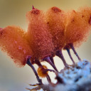
The fungus is cream-beige overall with carmel-brown veil that splits into scales like a Lepiota does! The stipe is white-pruinose but when touched or with age it is peach-brown, The flesh at the base is dark brown, lighter above. The ring is ascending, white above, peach-brown below
Spores in groups of 2-8 (now this is a Lepiota-thing!) tiny, difficult to explain the shape vut I would say elliptical with one end slightly more flattened than the other, but not distinctly so. They do not stain well with COngo red. Oil body and apiculum present
(3.5) 3.8 - 4.4 (4.6) × (2.1) 2.4 - 2.8 (3) µm
Q = (1.3) 1.5 - 1.7 (1.9) ; N = 35
Me = 4 × 2.6 µm; Qe = 1.6
Cheilocystidia quite frequent, grouped together and clavate to sphaeropedunculate, smallish,
15.74 − 27.39 x 5.27 − 9.7 µm (average 20 x 7 um)
basidia not much in good shape, but 4-sporous, small, with 2.8um long needle-thin sterigmata
The pileus skin is a trichoderm but the veil above is a hymenoderm of sphaeropedunculate shaped elements, quite similar to the cheilocystida, but remarkably larger - 36.9 x 14.1 µm (range: 26.11−42.84 x 11.56−16.36 µm)
Section 2.5-4.2cm high and cap 1.5-2.9 cm wide
Further details on the images
-
Dear Michael,
No problem, we are here to discuss, share opinions, learn and after all have fun communicating with real people rather than stuck in some AI app or screen. Thanks for the time to reply and have interest in my finding, and same goes to Oehrling, if he is still wants to talk to me after disappointing him a bit with the spore-stain
For now I place it as L. leucothites unless a better option arises, but as you said it looks alike and spore measurements are within the range, although from the literature shown, the range is quite wide!
Cheers!!!
-
Spore sizes of L. leucothites found on online literature
First nature: 7-9 x 4.5-5µm
University British Columbia: 7–10.0 x 5.0–7.0 µm
Mushroom expert: 8-11 x 5-6.5 µm
I also found a twin brother of my specimen, smallish with ring hanging on the rim of the pileus and no traces on the stipe!!!
Leucoagaricus leucothites (Vittad.) Wasser 1977 media - Encyclopedia of Life
-
New measurements in water.
9.27 5.77
7.93 5.18
7.23 5.45
8.55 5.08
8.79 6.25
8.28 6.04
7.83 5.36
8.83 6.05
8.00 5.91
7.20 5.37
7.55 5.16
6.45 5.40
8.78 5.60
8.55 5.40
8.82 6.03
6.84 5.49
8.31 5.06
7.36 5.59
8.99 5.30
8.77 6.24
7.45 6.05
7.83 5.60
8.12 5.73
8.05 5.91
7.90 5.72
7.36 4.88
8.16 5.76
7.47 5.29
7.57 5.68
7.77 5.58
6.90 5.00
8.04 5.27
7.38 5.25
7.63 5.34
8.34 5.95
8.28 5.59
7.49 5.46
8.57 5.78
7.99 5.94
9.55 5.46
7.89 5.45
8.94 5.73
8.51 5.65
8.71 5.68
7.67 4.88
(6.5) 7.2 - 8.8 (9.6) × (4.9) 5.1 - 6 (6.2) µm
Q = (1.2) 1.3 - 1.6 (1.7) ; N = 45
Me = 8 × 5.6 µm ; Qe = 1.4
-
Deeply sorry for not mentioning it - in many images the spores are stained in Congo Red (and also some post editing of the images to increase the contrast and make the bg whitish) and a few in Lugol's iodine to show their dextrinoid character typical of Leucoagaricus.
-
Some comparisons with Leucoagaricus cinerascens hold well.
https://www.mycocharentes.fr/pdf1/1331.pdf
-
Dear friends, I need your advice on this nice Leucoagaricus I found in a park under Pinus halapensis (and Eucalyptus) on calcareous soil. A rather robust basidiocarp, white/beige with a rather pale brown or coffee-milk pileus which in old specimens cracks into large scales. Spores dextrinoid, almond-ovate with rounded end and an indistinct or absent germ. pore (not sure about it to be honest, but seems not evident). The cheilocystidia are quite narrow and many are subcapitate or ventricose. The pilleipellis was also examined and terminal hyphae observed (see images). I am undecided about a few species such as L. leucothites? (must be white and has larger cheilocystidia!) L. wichanskyi (germ pore should be absent?) or L. cinerascens (cheilos are wide?) but I can't make up my mind.
Pileus up to 7cm wide
Spores dextrinoid 8.5 − 12.9 x 5.3 − 7.1 µm (10 x 6 um) / Q-factor: 1.4-2.0 (1.6)
Cheilocystidia subcapitate or ventricose 31.1 − 55.0 µm x 6.2 − 11.4 µm ( 42.28 9.14 µm)
-
Images attached here
-
Dear Oehrling and Michael, thank you for your replies.
Ring or not, I am not sure because I do not see a scar on the stem, but you could be right.
I reckon microscopy is required at this stage and I have marked that I have an exsiccatum for SM-552. I report findings over the weekend.
Thank you
LG / FG
Stephen
So finally I had time to examine the spores and gills of an exsiccatum which was not in very good shape (larvae destroyed physically part of the specimen. Here are some notes:
The gills turned beige-brown with age
Spore print white
Spore deposit/production abundant
Cystidia generally absent (but check image of a possible finding of a cheilocystidium)
spores broadly ovate or shield like with apiculum and distinct germ pore
Sizes:
(7.5) 7.8 - 9.3 (10.2) × (5) 5.3 - 6.1 (6.6) µm //
Me = 8.5 × 5.8 µm; Qe = 1.5 ; Ve = 147 µm3
Ring may be present and got detached and hanging at the rim during basidiocarp growth
I am thinking on Leucoagaricus, specifically L. leucothites ?
>I cannot upload pics!!!<
-
.... wanted to add that I bought this
The lowest temperature is 40C and unfortunately my thermometer (external metal thermometer) indicates 44C eith lid closed. Removing the lid and raising some compartments, I get around 36-40C. Hope this is fine for molecular testing and cell conservation for microscopy.
I wonder if anyone uses this too
-
This topic is of interest also to me. At the moment I have been struggling in finding a cheap or free image stacking software mostly for myxo but also for micro-images and small mushrooms. Any suggestions ?
Question 2 is that I have an SLR and I can have an adapter so as to fit in the microscope trinocular head (an old 1980s AxioLAB Zeiss). Do you have experience if photos taken this way produce good quality images. I am currently taking pictures of my eyepiece from an expensive point and shoot camera. Its 4x optical zoom enlarges the image nicely and it is very fast.
With the SLR the shooting is a bit slow procedure as I assume one has to refocus and adjust settings between live view and switching to shooting mode. Also I loose the ocular micrometer option.
-
Dear Oehrling and Michael, thank you for your replies.
Ring or not, I am not sure because I do not see a scar on the stem, but you could be right.
I reckon microscopy is required at this stage and I have marked that I have an exsiccatum for SM-552. I report findings over the weekend.
Thank you
LG / FG
Stephen
-
I am trying to identify this species found under carob trees and some pine needles from a tree further apart, on damp humus. No partial veil and I don't see a vulva. 26/11/2020
-
Thanks, will do over the weekend.
spores, cheilo/pleuro, and pileipellis.
-
Thank you, we have the same thoughts on this. I'll check the micro shortly. I was wondering if it is too small for L. subincarnata?
Thanks for replying
-
I have another Lepiota to show you. No microscopy for the time-being but specimen are being dried. Three specimens growing under oak tree (Quercus ilex) although Pine trees are also present not far. Specimen small, around 3cm tall with a cap about 2cm. Ring present but deteriorated with time. From my understanding, the stipe darkens to reddish brown with age that at one point when at home, I thought I might have gathered two different species, but on seeing photos in situ I believe they are all the same species. Margin pleated. Very elegant the tufts of veilar remains, toffee-brown in colour. Your opinions are welcomed on how to help to get an identification, unless molecular sequencein is the only way forward. Thanks!
-
Hello, rainy and cold season started in Malta and the mushrooms are punctual.
I was lucky to find something quite rare (for my eyes!) a small Lepiota with nice pattern of veilar remnants on the cap. I have dried a specimen for later microscopy, but at the moment I wish to have some clues. The fungus was about 2.5 cm tall and a cap of about 2 cm across. Scent indistinct or faint. It was growing in a mixed aforested area in a valley (sem-riparian) and the two trees present were Quercus ilex and Pinus halepensis. I have got an idea of L. subincarnata or because it is small, L. echinella.
I can carry microscopy after 5 days because I am away from my home Lab.
-
It has been decided at this hour during a meeting by JONEF members and we updated the deadline during this meeting to 10th Dec. It will be shown online soon, too.
Tnx
-
Dear myco friends,
I am part of a project called Jonef which is building a report for the conservation and protection of fungi based on two questionnaires.
Info on the project can be found here
Project JoNeF – Joint Network for wild Fungi | Network per la diversità micologicaThe direct links for these questions are found here.
PART 1
Füllen Sie bitte dieses Formular ausPART 2
Ausfüllen | JoNeF Questionnaire-base Survey (Part II)Deadline 10 December.
It is an easy online data input.
Your participation is welcomed and important especially because we lack data from germany, poland (and spain).
Thank you
-
This is Biscogniauxia mediterranea on dead Quercus ilex tree.
-
Hello Steve,
so far I think the new system is even better than the old one!
But I understand what you mean.
By the way: Fuligo was not placed with Diderma but Mucilago with Didymium, but I could well imagine that Fuligo will soon belong to Physarum.
Kind regards
Noah
Yes, sorry - bad memory - I meant that combination -> Mucilago into Didymium
-
Badhamia foliicolaHermosa especie de myxomycete con un capilicio delicado y tridimensional, parte inferior de la esporoteca oscura por transparentarse las esporas de su…www.flickr.comOccurrence Detail 3859253320Global Biodiversity Information Facility. Free and Open Access to Biodiversity Data.www.gbif.org
-
For a long time the study of myxomycetes was my favourite because the classification and keys and genera were quite straightforward and there was not much interference from genetic disruptions. Hope that more or less, things in slime moulds remain like this. Having Fulgido absorbed in Diderma is acceptable but let us see what happens in Badhamia and Physarum and if the basics we learnt have to be erased with new molecular concepts that not always make straight sense to a sensual brain!