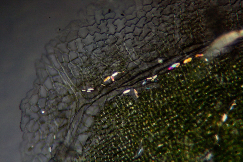
I had a lot of fun microscoping leaves, trying stains, polarisation etc. It begun when I needed to distinguish between Fraxinus excelsior and F. pennsylvanica on base of last year's skeletized leaves (Hymenoscyphus fraxineus + H. albidus grow on both species, but H. pusillus only on F. pennsylvanica). Fortunately, F. pennsylvanica is easily distinguished by the presence of large prismatic crystals along leaf veins. Here they are on a fresh leaf (native preparation, tangential section, polarised light), and on on old skeletized vein occupied by a Hymenoscyphus (piece of the vein squashed in KOH, polarized light):


Another task was to distinguish leaves of Prunus laurocerasus from Ilex aquifolium. The latter can be pretty variable, having all shapes from toothed to entire margin, which resemble the false laurel. Here is P. laurocerasus main vein in polarised light, slightly stained with cresyl blue:

Some trees can be distinguished not only by wood anatomy, but also by petiole anatomy - the vein bundles often have a genus-specific pattern. But this was the first time ever I looked into microscope and saw something smiling back at me (Sorbus aucuparia, water, in UV).


